
Skip to content

Our care

Specialist eye care,
explained clearly.

Explore diagnosis, treatment, surgery and vision correction across Mungale’s specialist areas of eye care.

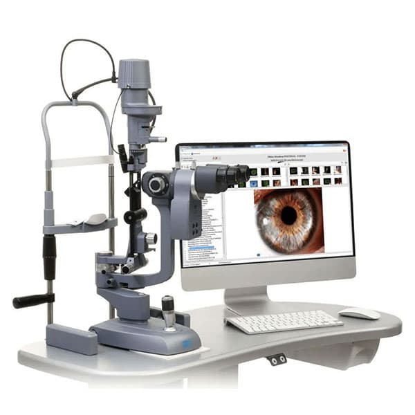
LED CSO slit lamp with imaging, showing an eye on the monitor
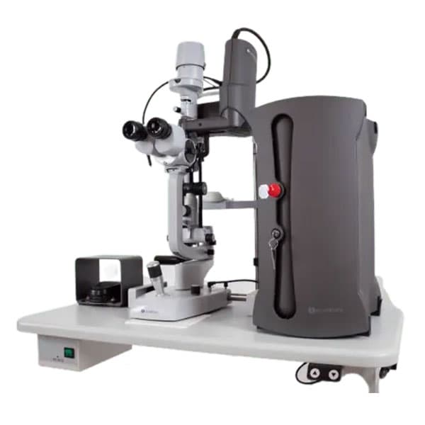
YAG laser machine used for peripheral iridotomy

LED slit-lamp imaging & YAG laser — diagnosis to treatment.

Explore by area of care

010203040506Cornea Evaluation · 01 / 06

Treatment atlas

Six areas of care, each with its own chapter. Scan the summaries — open the details only where you want them.

Diagnosis

Cornea Evaluation

Handy pachymeter — corneal thickness measurement

Seven specialist instruments map the cornea in depth — its shape, thickness, tear film and cell health — before any treatment is planned.

  1. Slit-lamp imaging
  2. Pachymetry
  3. Dry-eye analysis (IDRA)
  4. Specular microscopy
  5. Specialty lenses
  6. ANTERION tomography

In detail — cornea evaluation

Slit lamp with brilliant LED illumination which allows simultaneous visualisation by both doctors and relatives of the patient. It has imaging systems that will capture photos and real time videos of the eye. It helps the Doctors to see the minutest details of the patient’s eye, which includes all the parts of the eye. It is the gold standard for eye examination.

Pachymetry is a procedure to measure the corneal thickness of our eyes. This can be done in two ways: Optical and Ultrasonic of which the ultrasonic measurement is slightly more accurate. This is required in conditions of glaucoma and cornea.

The IDRA is a non-invasive, user-friendly ophthalmic device that analyzes tear film layers, diagnoses dry eye disease, offers automated lipid analysis, and provides 3D demonstrations with documented results and treatment options.

Specular microscopy is a photographic technique that allows you to visualize and analyze the corneal endothelium’s size, shape and population of the endothelial cells. It is useful in conditions like Fuch’s endothelial dystrophy, pseudophakic bullous keratopathy.

These are specialized contact lenses which provide excellent visual acuity in cases of Keratoconus.

Scleral contact lenses are large-diameter, gas-permeable lenses designed for individuals with keratoconus, dry eyes, Stevens-Johnson syndrome, or irregular corneas.

The ANTERION is an advanced, OCT-based device specifically designed for anterior segment imaging, including corneal tomography. It provides high-resolution cross-sectional images of the cornea, as well as detailed measurements of corneal thickness, curvature, and elevation. This helps in diagnosing and managing corneal diseases like keratoconus and planning procedures such as refractive surgeries. The device’s precision allows for a comprehensive evaluation of the cornea, ensuring accurate and reliable assessments.

This is the complete care information published by Mungale Eye Hospital for cornea evaluation. Anything you need beyond it is best discussed in person.

End of chapter 1

Diagnosis

Glaucoma Evaluation

Fundus imaging — the retina, photographed

Eye pressure, drainage angles, visual fields and optic-nerve imaging combine into one complete glaucoma work-up.

  1. Tonometry
  2. Gonioscopy
  3. Visual-field analysis
  4. Fundus photography
  5. Pachymetry
  6. OCT

In detail — glaucoma evaluation

At MEH we have tonometers as shown below, which measures the eye pressure.

The most ideal way to measure intra-ocular pressure gives accurate readings always.

Ideal tonometer for immobile pts/old patients/pediatric pts to be examined under general anesthesia.

For scarred corneas, edematous corneas, and irregular astigmatism following keratoplasty.

Gonioscopy is performed during the eye exam to evaluate the internal drainage system of the eye, also referred to as the anterior chamber angle. Gonioscopy lenses help us to differentiate different types of glaucomas which in turn helps us to treat glaucomas appropriately.

The ZEISS Humphrey Field Analyzer 3 (HFA3) maps visual fields, detects early vision loss, and aids in treatment planning for conditions like glaucoma.

Fundus photography is taking the image of the retina of the eye with a fundus camera. Fundus photography is important for diagnosing and treating various posterior segments and other ocular diseases. The illumination and reflectance of the retina occur through the common optical path, i.e., the pupil.

iCare IC100 is a reliable choice for all eye care professionals. With this tonometer, you can measure the intraocular pressure of patients in a sitting or standing position. Optometrists and ophthalmologists have successfully used IC100 for routine IOP measurement and glaucoma screenings, as measuring has never been more accessible – just load, align and measure.

It measures corneal thickness. Corneal thickness affects glaucoma management; hence, measurement of corneal thickness is very important.

Peripheral vision is affected in glaucoma. Hence in the initial part of the disease the patient is unaware of this and is symptom free. Measurement of peripheral vision helps us in quantifying the disease which helps in further management of the disease. The test assesses the functional damage to the eye. The visual field exam takes about half an hour. Each eye is tested separately. The visual field needs to be repeated at least every year.

OCT is very useful in assessing the structural damage to the eye. It helps in identifying early glaucoma cases.

This is the complete care information published by Mungale Eye Hospital for glaucoma evaluation. Anything you need beyond it is best discussed in person.

End of chapter 2

Treatment & Surgery

Corneal Treatments

Transplantation — clinical illustration

Medical and surgical care for keratoconus, infection, injury and surface disease — including partial and full corneal transplantation.

  1. Keratoconus
  2. Corneal infection
  3. Transplantation
  4. Chemical burns
  5. Dry eye & allergy
  6. Stevens–Johnson syndrome

In detail — corneal treatments

Keratoconus is a progressive eye disease where the cornea thins and bulges into a cone shape, distorting vision. Treatments for progressive keratoconus include: corneal collagen cross-linking (C3R); scleral and semi-scleral lenses; corneal transplant.

Eye infections can be caused by viruses, bacteria, or fungi. A microbiology test (scraping) helps determine whether it’s bacterial or fungal, guiding treatment.

Corneal transplantation involves replacing a diseased cornea with a healthy donor cornea. Indications include infection, keratoconus, or post-surgery complications. Newer techniques allow for partial corneal replacement: Deep anterior lamellar keratoplasty (DALK) replaces anterior layers of the cornea; Descemet’s stripping endothelial keratoplasty (DSEK) replaces Descemet’s layer; Penetrating keratoplasty (PK) replaces all five layers of the cornea.

Patients with chemical eye injuries require emergency medical care. Treatment may involve amniotic membrane transplantation, tenonplasty, or tarsorrhaphy. Further treatment could include conjunctival autografts, Simple limbal epithelial transplant (SLET), penetrating keratoplasty, keratoprosthesis for vision restoration.

Evaluation of dry eye involves assessing tear production and evaporation.

Allergic eye symptoms can be easily identified and may contribute to vision loss if untreated.

A rare disorder affecting the skin and mucous membranes, often triggered by medications or infections. Treatment involves ocular lubrication and surveillance of infections. In the acute stage, amniotic membrane transplantation is performed, while in the chronic stage, mucous membrane grafts and scleral lenses are offered.

This is the complete care information published by Mungale Eye Hospital for corneal treatments. Anything you need beyond it is best discussed in person.

End of chapter 3

Treatment & Surgery

Glaucoma Treatments

YAG laser — peripheral iridotomy

Laser procedures and surgery that lower eye pressure — from SLT and YAG iridotomy to micro-invasive and valve techniques.

  1. SLT laser
  2. YAG iridotomy
  3. Conventional surgery
  4. MIGS
  5. Valve implants
  6. Congenital glaucoma

In detail — glaucoma treatments

SLT is useful for reducing intra-ocular pressure and helping patients maintain a life on minimum intraocular drops. This procedure is very safe and can be repeated. Success rate of this procedure is 90%.

We use a laser beam (the YAG laser) to make a microscopic hole in the iris (the colored part of the eye) so that the fluid in the eye flows through the hole and the eye pressure remains under control. It is a very safe and effective procedure to treat angle-closure glaucoma.

1) Conventional Surgery — Trabeculectomy with MMC (Mitomycin-C): a filtration surgery to lower intraocular pressure (IOP) by creating a drainage flap. 2) MIGS (Minimal Invasive Glaucoma Surgery) — GATT (Gonioscopy-Assisted Transluminal Trabeculotomy), where the trabecular meshwork is opened to improve outflow of fluid; KDB (Kahook Dual Blade), a surgical tool used to excise part of the trabecular meshwork; iStent, a tiny device implanted to create a pathway for fluid drainage and lower IOP. 3) Valve Surgeries — AGV (Ahmed Glaucoma Valve), a valve implanted to control IOP by regulating fluid drainage; ACP (Ahmed ClearPath), a variation of AGV with a tube designed for improved positioning; AADI (Aurolab Aqueous Drainage Implant), a cost-effective implant designed to regulate IOP. 4) Surgery for Congenital Glaucoma — Trabeculotomy + Trabeculectomy, a combined approach for more effective IOP control; Goniotomy, a surgical procedure to open the angle structures of the eye for better fluid outflow; Valve Surgeries, similar to adult procedures, designed for severe cases; GATT, also utilized for congenital cases to enhance outflow from the anterior chamber.

This is the complete care information published by Mungale Eye Hospital for glaucoma treatments. Anything you need beyond it is best discussed in person.

End of chapter 4

Treatment & Surgery

Cataract Surgery

Oertli CataRhex 3 — phaco platform

Cataract removal by phacoemulsification on two specialist platforms, with intraocular lens options to suit each eye.

  1. Ellips FXO phaco system
  2. Oertli CataRhex 3
  3. Intraocular lenses

In detail — cataract surgery

Ellips FXO AMO Phaco Machine (Sovereign Compact System With Advanced Ellips FXO Technology): MEH boasts of one of the advanced systems for phacoemulsification which helps remove cataract with high efficiency in matter of minutes. Additionally all types of intraocular lenses are offered at very reasonable prices.

The Oertli CataRhex 3 is a surgical platform used for cataract and glaucoma surgery. A compact and lightweight phaco machine with an integrated compressor for anterior vitrectomy. It has a control panel with bright displays, logical button arrangement, and a pivotable display panel. The CataRhex 3 can be used for both cataract and glaucoma surgery.

This is the complete care information published by Mungale Eye Hospital for cataract surgery. Anything you need beyond it is best discussed in person.

End of chapter 5

Vision Correction

Optical & Contact Lenses

The Mungale optical — frames on display

Spectacles and custom-fit contact lenses for everyday and complex vision needs, verified on the in-house lensometer.

  1. Spectacles & contact lenses
  2. Specialty keratoconus lenses
  3. Custom fitting
  4. GL7000 verification

In detail — optical & contact lenses

We offer our eye patients various types of spectacles and contact lenses. This includes rigid lenses, soft lenses, and cosmetic lenses to change eye color. In addition, specialty contact lenses for keratoconus which includes Rose K lenses, mini scleral lenses, and Boston scleral lenses (PROSE) are also provided. Our contact lenses are custom-fit for your eyes and vision.

The GL7000 also has a full-graphic LCD monitor that displays the measured values for both eyes at once. It comes with a high-speed thermal printer to quickly print out the measured data. It has the capability to verify the prescription of your Glasses Lens as well as your hard/soft Contact Lens.

This is the complete care information published by Mungale Eye Hospital for optical & contact lenses. Anything you need beyond it is best discussed in person.

End of chapter 6

Care around your treatment

The hospital, behind the treatment.

In-house pharmacy

At MEH we have an in-house pharmacy, where our eye patients easily buy medicines according to their treatment. Our staff is fully trained to explain and instill medicines in your eyes.

Well-equipped operation theatre

MEH has one of the largest eye operation theatres in Baroda city. It has two OT tables and 2 LED microscopes. Eye surgeries can be performed under General anesthesia which makes us equipped to perform pediatric eye surgeries, corneal tears, and emergency cases. We also have the class B sterilizer which allows us to autoclave all hollow tubings required for eye surgeries under strict safety and quality controls. In addition, an ETO sealing machine for ethylene oxide sterilization is also available. No compromise in ensuring sterility and safety for eye surgeries!

CallWhatsAppBook