Skip to content

Medically reviewed by Dr. Sachin Mungale, MS (Ophthalmology) · Glaucoma Fellowship, LVPEI · Gujarat Medical Council Reg. No. G-12281 · Reviewed on 3 October 2026

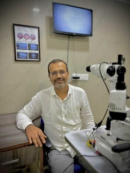
Dr. Sachin Mungale at the slit lamp

Dr. Sachin Mungale

MS (Ophthalmology) · Glaucoma Fellowship, LVPEI

Diagnosis — Glaucoma

Glaucoma Evaluation

Pressure, angles, fields and nerve imaging — a complete work-up that finds glaucoma early, often before symptoms.

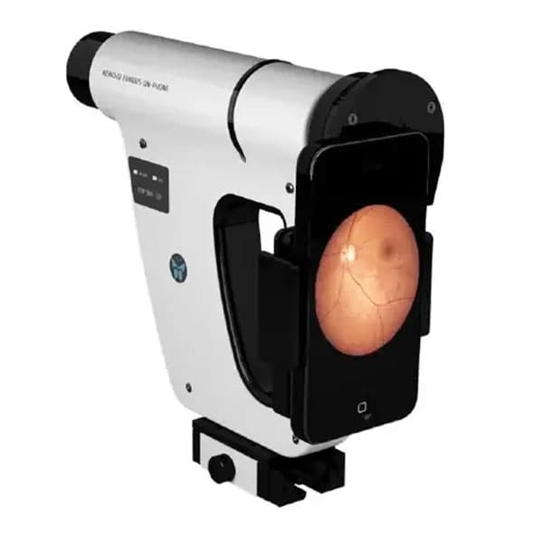
Handheld fundus camera displaying a retinal image
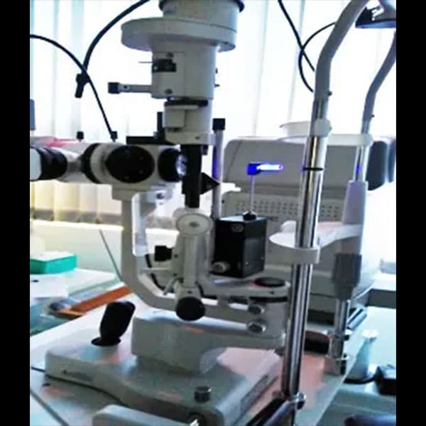
Slit-lamp examination at Mungale Eye Hospital

Fundus imagingthe retina, photographed

Glaucoma care · 5 groups

010203040506Glaucoma Evaluation

How glaucoma is evaluated

A complete work-up,
piece by piece.

Five groups of examination — pressure, angles, visual function, retinal structure and corneal measurement — each answering a different question about glaucoma.


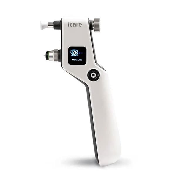
iCare IC100 tonometer for eye-pressure measurement

iCare tonometerclinical photograph

Eye Pressure

Four tonometers cover every eye — routine, immobile, paediatric, scarred and post-surgical corneas.

At MEH we have tonometers as shown below, which measures the eye pressure.

The most ideal way to measure intra-ocular pressure gives accurate readings always.

Ideal tonometer for immobile pts/old patients/pediatric pts to be examined under general anesthesia.

For scarred corneas, edematous corneas, and irregular astigmatism following keratoplasty.

iCare IC100 is a reliable choice for all eye care professionals. With this tonometer, you can measure the intraocular pressure of patients in a sitting or standing position.
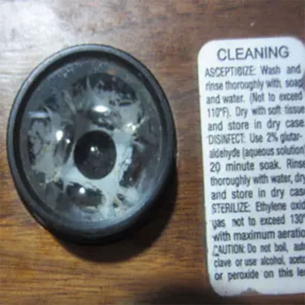
Gonioscopy lens used to examine the eye's drainage angle

Gonioscopy lensclinical photograph

Angle & Structure

Gonioscopy examines the eye's drainage system to tell glaucoma types apart — and treat each correctly.

Gonioscopy is performed during the eye exam to evaluate the internal drainage system of the eye, also referred to as the anterior chamber angle. Gonioscopy lenses help us to differentiate different types of glaucomas which in turn helps us to treat glaucomas appropriately.

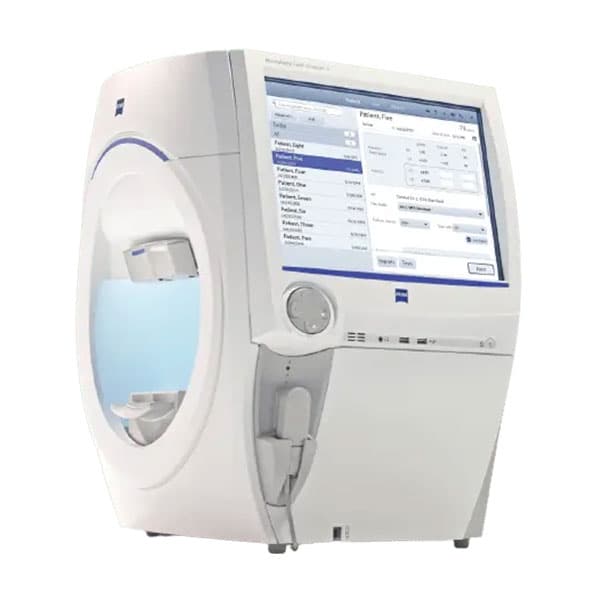
ZEISS Humphrey Field Analyzer 3 for visual-field mapping

Visual-field analyzerclinical photograph

Visual Function

Visual-field mapping quantifies what glaucoma silently takes — repeated at least every year.

The ZEISS Humphrey Field Analyzer 3 (HFA3) maps visual fields, detects early vision loss, and aids in treatment planning for conditions like glaucoma.

Peripheral vision is affected in glaucoma. Hence in the initial part of the disease the patient is unaware of this and is symptom free. Measurement of peripheral vision helps us in quantifying the disease which helps in further management of the disease. The visual field needs to be repeated at least every year.

Handheld fundus camera displaying a retinal image

Fundus imagingthe retina, photographed

Retinal Assessment

Fundus photography and OCT reveal structural damage — including early glaucoma.

Fundus photography is taking the image of the retina of the eye with a fundus camera. Fundus photography is important for diagnosing and treating various posterior segments and other ocular diseases.

OCT is very useful in assessing the structural damage to the eye. It helps in identifying early glaucoma cases.

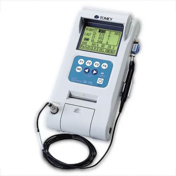
Tomey SP-100 handy pachymeter used to measure corneal thickness

Handy pachymetercorneal thickness measurement

Corneal Measurement

Corneal thickness changes glaucoma management — so it is measured, not assumed.

It measures corneal thickness. Corneal thickness affects glaucoma management; hence, measurement of corneal thickness is very important.

A closer look

The instruments, up close.

Fundus imaging — the retina, photographed

CallWhatsAppBook