
Medically reviewed by Dr. Sachin Mungale, MS (Ophthalmology) · Glaucoma Fellowship, LVPEI · Gujarat Medical Council Reg. No. G-12281 · Reviewed on 3 October 2026
Published 22 May 2026 · Updated 22 May 2026
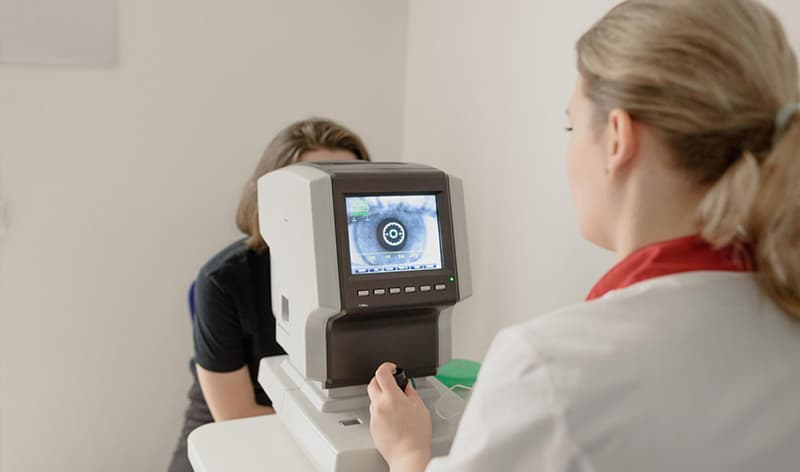
Progression measurement benchmarks in eye care are standardized metrics used to track the advancement of a chronic eye condition over time. Doctors establish a baseline for a patient’s vision and eye health using tools like OCT scans and visual field tests. They then compare subsequent measurements against this baseline to determine if the disease is stable or worsening, allowing for timely adjustments to treatment plans to preserve vision.
Why are these benchmarks important for your vision?
Progression benchmarks translate complex data from eye exams into a clear picture of your eye health trajectory. For chronic conditions like glaucoma or diabetic retinopathy, vision loss is often gradual and goes unnoticed by the patient until significant damage has occurred. These benchmarks act as an early warning system. By tracking subtle changes in intraocular pressure (IOP), retinal thickness, or peripheral vision, your ophthalmologist can intervene before you experience noticeable symptoms. This data-driven approach ensures that treatment decisions are based on objective evidence, not guesswork, which is critical for preserving sight long-term.
How are benchmarks used for different eye conditions?
Different eye diseases affect the eye in unique ways, requiring distinct benchmarks and measurement tools for effective monitoring. The approach for tracking pressure changes in glaucoma is very different from monitoring retinal changes in macular degeneration.
| Condition | Primary Benchmark | Key Measurement Tool | Goal of Monitoring |
|---|---|---|---|
| Glaucoma | Intraocular Pressure (IOP) & Visual Field Loss | Tonometry & Perimetry (Visual Field Test) | Prevent optic nerve damage by keeping IOP within a target range (e.g., below 18-20 mmHg). |
| Myopia (in Children) | Axial Length & Refractive Error | Biometry & Autorefractor | Slow the elongation of the eyeball to reduce the risk of high myopia and associated diseases later in life. |
| Age-Related Macular Degeneration (AMD) | Drusen Size/Volume & Retinal Fluid | OCT Scan & Fundus Photography | Detect the transition from dry to wet AMD and monitor treatment effectiveness by tracking retinal swelling. |
| Diabetic Retinopathy | Retinal Thickening & New Blood Vessel Growth | OCT Scan & Fluorescein Angiography | Identify and treat macular edema or proliferative retinopathy before it causes irreversible vision loss. |
What are the common measurement tools involved?
To establish and track benchmarks, eye care specialists rely on precise diagnostic technology. These tools provide the objective data needed to monitor your condition accurately.
Optical Coherence Tomography (OCT)
An OCT scan is a non-invasive imaging test that uses light waves to take cross-section pictures of your retina. It allows your doctor to measure the thickness of the retina and its layers with micrometer-level precision. This is essential for monitoring retinal diseases like AMD and diabetic retinopathy, as it can detect fluid leakage or thinning of the nerve fiber layer in glaucoma long before it impacts vision.
Visual Field Testing (Perimetry)
This test maps your complete field of vision, including your central and peripheral (side) vision. During the test, you’ll be asked to identify flashes of light. It is a fundamental benchmark for glaucoma because it can detect blind spots caused by optic nerve damage. An expanding or deepening blind spot over a series of tests is a clear indicator of disease progression.
Tonometry measures the pressure inside your eye, known as intraocular pressure (IOP). While a single high reading is a risk factor, the more critical benchmark is the pattern of IOP over time. A change of more than 3-4 mmHg from the established baseline may be considered significant and prompt a change in treatment for a glaucoma patient.
Operational Authority Block: Your Checklist for Discussing Progression with Your Doctor
Use this checklist to have a more informed conversation with your eye care provider. Understanding your personal benchmarks empowers you to be an active participant in your care.
When is monitoring progression most critical?
This type of detailed monitoring is not for everyone. It is most critical for individuals diagnosed with chronic, progressive eye diseases. This includes patients with glaucoma, age-related macular degeneration (AMD), diabetic retinopathy, and children with rapidly advancing myopia. It is also essential for individuals with a strong family history of these conditions or those identified as being at high risk. For those with stable vision and no underlying conditions, a standard comprehensive eye exam is typically sufficient.
How to find a specialist for eye care monitoring in Vadodara?
For conditions requiring progression monitoring, you need an ophthalmologist or a specialized eye care center with the right diagnostic equipment. In Vadodara, look for clinics that explicitly mention services for glaucoma management, retinal diseases, or pediatric myopia control. When booking an appointment, you can ask if they have OCT and visual field testing capabilities. Reputable local hospitals and specialized clinics, such as the Mungale eye care facilities, are equipped to establish and monitor these critical health benchmarks.
Concerned about changes in your vision or diagnosed with glaucoma, diabetic retinopathy, AMD, or myopia? Book an eye evaluation at Mungale Eye Hospital in Vadodara for proper monitoring and timely care.
Frequently Asked Questions
While it varies per patient, a consistent change of over 3-4 mmHg from your established baseline is often considered significant by ophthalmologists. The overall trend across multiple visits is more important than a single reading, as IOP can fluctuate throughout the day.
The frequency depends on the condition and its severity. A stable glaucoma patient might be monitored every 6-12 months, while someone with active wet AMD may require scans every 4-6 weeks. Your doctor will determine the appropriate schedule based on your individual risk profile.
Yes, there are widely accepted clinical guidelines, like the Early Treatment Diabetic Retinopathy Study (ETDRS) severity scale. Doctors use this framework, along with OCT scans and eye photos, to classify the disease stage and determine when treatments like laser therapy or injections are necessary.
For some conditions, yes. In glaucoma, managing blood pressure and adhering to medication schedules can help stabilize IOP. For diabetic retinopathy, strict blood sugar and blood pressure control is the most effective way to slow progression and improve benchmarks.
Most tests, like OCT scans and tonometry, require no special preparation. For a visual field test, it is helpful to be well-rested to ensure concentration. If you are having a test that requires pupil dilation, such as detailed retinal photography, you will need to arrange for someone to drive you home.