
Written by the Mungale Eye Hospital team
Medically reviewed by Dr. Meeta Mungale, MBBS, MS (Ophthalmology), DNB · Fellowship in Cornea & Anterior Segment, L V Prasad Eye Institute, Hyderabad · Gujarat Medical Council Reg. No. G-39262 · Reviewed on 5 October 2026
Published 5 October 2026
Part of our guide: Keratoconus: Symptoms, Causes, Diagnosis & Treatment
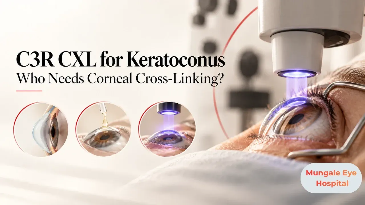
Corneal cross-linking, known as CXL or C3R (corneal collagen cross-linking with riboflavin), is a treatment for keratoconus. It strengthens the cornea by creating new bonds between collagen fibres. The main goal is to stop or slow the progression of keratoconus, not to restore perfect vision.
How Does Cross-Linking Work?
The cornea is made of collagen fibres. In keratoconus, these fibres are weak, allowing the cornea to thin and bulge. Riboflavin drops soak into the cornea and, when exposed to controlled ultraviolet A light, create additional bonds between collagen fibres, making the cornea stiffer and more resistant to further bulging.
Who Is a Good Candidate?
Patients with very thin corneas, severe scarring, active infection or certain other eye conditions may not be suitable or may need modified techniques.
What Are the Types of CXL?
Epithelium-off (standard)
The thin surface layer, the epithelium, is gently removed so riboflavin penetrates well. This is the most established method.
Epithelium-on (transepithelial)
The surface layer is left intact. Recovery is faster, but effectiveness may vary by technique.
Accelerated protocols
Higher-intensity light reduces treatment time. Your doctor chooses the method.
What Happens During the Procedure?
Numbing drops are applied. In epi-off CXL, the epithelium is removed. Riboflavin drops are applied for a period, then the eye is exposed to UVA light while drops continue. A bandage contact lens is placed at the end. The procedure usually takes about an hour.
What Is Recovery Like?
Discomfort, light sensitivity and watering are common for a few days after epi-off CXL. Vision may be blurry for weeks and gradually improve. Antibiotic and anti-inflammatory drops are used. Follow-up scans check stability. Final effect is assessed over months.
What Can CXL Not Do?
CXL does not cure keratoconus or reverse existing cone shape significantly. Some patients have mild flattening and small vision improvement, but most still need glasses, contact lenses or scleral lenses. In advanced cases, other procedures may be needed.
What Are the Risks?
Risks include infection, delayed healing, corneal haze, scarring and, rarely, failure to halt progression. Careful patient selection and follow-up reduce risks.
Key Takeaways
Final Thoughts
Cross-linking is one of the most important advances in keratoconus care because it can preserve vision long term when done early. Dr. Meeta Mungale and the team at Mungale Eye Hospital in Vadodara offer keratoconus assessment and cross-linking.
Frequently Asked Questions
C3R or CXL is corneal collagen cross-linking with riboflavin. It uses riboflavin drops and ultraviolet light to strengthen the cornea and slow or stop keratoconus progression.
The main goal is to stabilise the cornea, not to improve vision. Some patients notice mild improvement, but most still need glasses, contact lenses or other treatment for clear vision.
People with documented progressive keratoconus, especially teenagers and young adults with adequate corneal thickness, are typical candidates. Your eye doctor decides after serial scans.
The procedure is done with numbing drops. Afterward, discomfort, watering and light sensitivity are common for a few days after epithelium-off treatment, managed with drops and pain relief.
Cross-linking successfully stabilises keratoconus in most patients, but progression can occasionally continue. Regular follow-up scans monitor the cornea.
Sources and further reading
Reviewed by
MBBS, MS (Ophthalmology), DNB · Fellowship in Cornea & Anterior Segment, L V Prasad Eye Institute, Hyderabad
Gujarat Medical Council Reg. No. G-39262
Book a consultation
2nd Floor, Vinraj Plaza, opp. Government Press, Kothi Road, Anandpura, Vadodara, Gujarat 390001
Mon - Sat, 09:00 AM - 08:00 PM · Sunday closed
Related Articles
Start with a conversation.
Appointments
+91 81400 50055