
Medically reviewed by Dr. Meeta Mungale, MS (Ophthalmology), DNB · Cornea Fellowship, LVPEI · Gujarat Medical Council Reg. No. G-39262 · Reviewed on 3 October 2026
Published 28 September 2026 · Updated 28 September 2026
The cornea is supposed to be a smooth, evenly curved dome sitting at the front of the eye. In keratoconus, part of it thins out and starts pushing forward into a cone shape instead. That change bends incoming light unevenly, which is why vision in keratoconus often looks blurred, streaked, or doubled in a way that a fresh glasses prescription doesn't seem to fix. It usually starts showing up in the teenage years or early twenties and tends to develop over several years before settling down.
Quick Answer
Keratoconus happens when the cornea progressively thins and bulges outward, distorting how light focuses inside the eye. Doctors diagnose it using corneal topography, a scan that maps the exact curve of the corneal surface, sometimes alongside a thickness measurement called pachymetry. Treatment depends on the stage: contact lenses (often rigid or scleral lenses once glasses stop working), corneal cross-linking (C3R) to slow or stop further change, and, for the more advanced cases, a corneal transplant. Nothing currently reverses the shape once it's changed, but with the right combination of monitoring and treatment, most people keep workable vision for the long run.
Why It Matters
People often notice keratoconus indirectly, before they know what it is. Maybe the optician keeps updating the prescription every few months. Maybe a new pair of glasses corrects things on paper but the vision still feels smeared, like looking through a slightly warped window. Both of those are worth mentioning at an eye exam, because keratoconus tends to respond better to treatment while the cornea is still comparatively stable.
None of this needs to feel like an emergency. Most people diagnosed with keratoconus go on living completely ordinary lives once their vision is corrected properly, and treatment has genuinely improved over the past couple of decades, cross-linking especially. What follows is a plain explanation of what's actually happening inside the eye, how doctors work out what stage it's at, and what treatment realistically looks like depending on where things stand.
What Is Keratoconus?
Picture the cornea as similar in shape to a contact lens: curved, symmetrical, and structurally consistent across its whole surface. Together with the lens inside the eye, it bends light so it lands precisely on the retina. In keratoconus, one section of that structure becomes weaker than the tissue around it. Ordinary internal eye pressure then pushes on that weak spot over time, and it slowly bulges forward into a cone, usually a little off-center rather than dead in the middle.
Once the surface stops being uniformly curved, light no longer focuses to a clean point. It scatters instead. That's where the hallmark symptoms come from: blurring, halos or streaks around light sources, and a specific kind of visual distortion called irregular astigmatism, which behaves quite differently from the more common astigmatism found in otherwise normal corneas.
Both eyes are usually involved, though it's fairly typical for one eye to run well ahead of the other, sometimes by years.
Causes and Risk Factors
There isn't a single identified cause. Most of what's understood points to a mix of inherited and environmental factors that weaken the collagen structure holding the cornea's shape together.
A few things raise the odds:
If several of these apply to you, particularly a family history combined with a habit of rubbing your eyes, it's worth flagging to an eye doctor even before anything feels wrong, just so a baseline scan is on file.
Symptoms and When to See a Doctor
Early on, keratoconus can be easy to miss or brush off as needing new glasses. What tends to show up:
Worth booking an exam if: your glasses prescription has changed more than once recently, your vision still feels smudged or ghosted even after an update, or you find yourself squinting constantly just to bring things into focus. A comprehensive exam that includes corneal imaging is the right next step.
One thing worth knowing about, even though it's uncommon: in advanced keratoconus, the inner lining of the cornea can occasionally develop a sudden tear, letting fluid rush into the tissue. This causes sudden eye pain, redness, and a sharp drop in vision, a condition called acute corneal hydrops. It's rare, but if it happens, it needs same-day attention rather than a routine appointment slot.
How Keratoconus Is Diagnosed
Standard vision charts won't catch keratoconus reliably, since the problem is the corneal shape itself rather than general clarity. Diagnosis typically draws on a few tools:
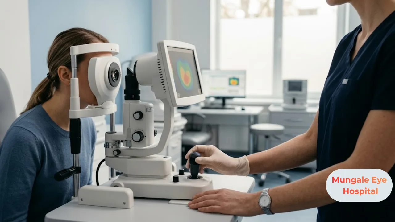
Corneal topography does most of the heavy lifting. It maps the curve of the entire corneal surface and generates a color map showing exactly where the steepening is happening. This can flag keratoconus well before it's visible during a routine exam.
Pachymetry measures how thick the cornea is at different points. Since thinning is central to the disease, this gives doctors a number to track over repeat visits.
A slit-lamp exam lets the doctor look closely at the cornea under magnification, checking for specific physical signs associated with keratoconus.
Refraction testing quantifies how much irregular astigmatism is present and gives another data point to compare visit over visit.
For a new diagnosis, it's common to repeat topography and pachymetry every few months rather than making a treatment call off a single scan. Whether the cornea is holding steady or actively changing shapes the whole treatment conversation that follows.
Treatment Options
What gets recommended depends on two things mainly: how far along the condition is, and whether it's currently progressing or has settled. Broadly, there are three paths.
Glasses and Contact Lenses
Early keratoconus sometimes still responds to glasses or ordinary soft contacts. As the corneal surface gets more irregular, though, these tend to stop cutting it, because they can't compensate for a surface that isn't uniformly curved anymore.
At that point, specialty lenses usually take over:
None of these lenses stop the cornea from continuing to change underneath them. They correct vision, not the underlying process.
Corneal Collagen Cross-Linking (C3R/CXL)
Cross-linking is the main treatment aimed at halting progression rather than just correcting how you see. Riboflavin (vitamin B2) drops are applied to the cornea, followed by a carefully controlled dose of UV light. This reaction essentially strengthens the bonds between collagen fibers, giving the cornea more structural rigidity than it had before.
It's worth being clear about what cross-linking does and doesn't do. It doesn't undo the cone that's already formed. What it aims for is stopping things from getting worse. It tends to work best in people whose scans show active progression, and earlier intervention, before much thinning has occurred, generally gives better odds of holding the shape steady.
Intracorneal Ring Segments
For select patients, small curved implants can be placed inside the cornea to help flatten the cone somewhat, which can also make contact lenses fit and perform better. This isn't a routine first-line step; it's considered case by case.
When a Corneal Transplant Comes Into the Picture
A smaller group of patients reach a point where the cornea is too thin, too scarred, or too irregular for lenses or cross-linking to give usable vision anymore. That's when a transplant gets discussed. Depending on which corneal layers are involved, this might mean replacing the full thickness of the cornea, or only certain layers, an approach known as lamellar keratoplasty. This decision comes after the other options have genuinely been tried and is based on detailed imaging plus how much functional vision the person actually needs day to day.
What to Expect During Cross-Linking
It's done as an outpatient procedure. Numbing drops go in first, then riboflavin solution saturates the cornea, followed by a set period of controlled UV exposure. Most of this wraps up in under an hour. Afterward, a soft bandage contact lens is typically placed to protect the healing surface.
Because the outer layer of the cornea is involved, some discomfort, light sensitivity, and blurry vision in the first few days is normal. Vision generally settles over the following weeks as healing progresses.
Recovery and Aftercare
After cross-linking, patients are usually told to:
For anyone moving into rigid or scleral lenses, getting used to how they feel takes a bit of time, and follow-ups are usually needed to refine the fit as the cornea's shape gets reassessed.
Risks and Limitations
Cross-linking is generally well tolerated. That said, some temporary haze in the cornea during healing is common, and in a small number of cases, healing runs slower than expected or an infection develops, which is exactly why follow-up visits aren't optional. It also won't restore a shape that's already changed. This is a stabilizing treatment, not a correction, so most people still need glasses or specialty lenses afterward.
Scleral and RGP lenses come with their own adjustment period and require careful handling and hygiene, much like any contact lens.
Cost and Insurance Considerations
Managing keratoconus isn't a single line-item cost. It depends on the stage of the condition, what diagnostic imaging is needed, whether cross-linking gets recommended, and what type of lenses end up being the right fit. Each of these is priced separately, and the total varies from one patient to the next depending on that combination. The most useful thing to do is discuss your specific scan results, treatment plan, insurance coverage, and cashless options directly with the hospital's billing team once your evaluation is done.
Common Myths About Keratoconus
"Keratoconus always leads to blindness." For most people, it's manageable through lenses, cross-linking, or in rarer cases, a transplant. Complete vision loss isn't the typical outcome.
"Glasses can always fix it." Once the cornea gets irregular enough, glasses often can't do what specialty contact lenses can.
"Cross-linking makes vision better right away." Its main job is stopping progression. Any visual improvement tends to show up gradually, and it's a side effect of stabilization rather than the point of the procedure.
"If one eye looks fine, it'll stay fine." Keratoconus usually shows up in both eyes eventually, even when one is diagnosed or progresses well ahead of the other.
How to Choose an Eye Hospital or Specialist
Keratoconus care works best with continuity, since it involves repeat imaging over months or years just to see whether things are holding steady. Look for a center with in-house corneal topography and pachymetry, direct access to cross-linking if progression shows up, and an actual lens-fitting service for rigid or scleral lenses. Not every clinic offers all three. At Mungale Eye Hospital, corneal evaluation, cross-linking, and specialty lens fitting are all available under one roof, along with monitoring for glaucoma and cataract, which matters for the patients managing more than one eye condition at once.
Latest Developments
Cross-linking protocols keep getting refined, with variations in how riboflavin is applied and how UV exposure is timed, generally aimed at improving comfort and cutting down treatment time without sacrificing effectiveness. Corneal imaging has also gotten sharper, catching keratoconus at earlier stages, sometimes before a patient notices anything wrong, which opens the door to earlier treatment decisions.
Common Mistakes to Avoid
Best Practices for Managing Keratoconus
Expert Tips
If you have old prescriptions lying around, bring them to your first evaluation. A documented history of how your prescription has shifted over a few years gives an ophthalmologist real information to work with when assessing how quickly things might be moving.
Key Takeaways
Final Thoughts
For most people, keratoconus turns out to be manageable, especially when it's caught while the cornea is still fairly stable. Accurate imaging, cross-linking when there's active progression, and a good lens fit cover the majority of cases without ever needing surgery. If your glasses prescription keeps changing or your vision has that persistent smeared quality no update seems to fix, getting a corneal evaluation is a reasonable move, and doing it sooner tends to leave more options open later.
If any of this sounds familiar, a corneal evaluation at Mungale Eye Hospital can tell you whether keratoconus is present, what stage it's at, and which treatment path actually fits your situation.
FAQ SECTION
Not in the sense of reversing the corneal shape once it's changed, no. Cross-linking can stop or slow further change, and lenses (or, in advanced cases, a transplant) can restore usable vision. Most people manage the condition well over the long term even without the shape itself going back to normal.
Most commonly the teenage years through the mid-twenties, though earlier or later diagnoses do occur. Progression tends to slow down with age for a lot of people, which is part of why catching it early and monitoring it matters.
A family history raises the odds, but a large share of people diagnosed have no affected relatives at all. It's generally thought to come from a combination of genetic and environmental factors rather than one inherited cause.
Heavy, chronic eye rubbing is one of the more consistently linked risk factors, often tied to underlying allergies. Cutting back on rubbing and treating allergies is worth doing regardless of what treatment stage you're at.
Not really during the procedure itself, since numbing drops are used throughout. Some discomfort, light sensitivity, and blurred vision for a few days afterward is typical while the surface heals.
Early on, sometimes yes, soft lenses or glasses can still work. Once the cornea gets more irregular, specialty lenses like RGP or scleral lenses generally give clearer vision than standard contacts can manage.
That depends on whether your case is stable or actively progressing, which your ophthalmologist determines from topography and pachymetry results over time. Progressing cases tend to get watched more closely than stable ones.
Most people never do. It's generally reserved for the more advanced cases, where the cornea's become too thin, scarred, or irregular for lenses or cross-linking to provide workable vision anymore.