
Medically reviewed by Dr. Sachin Mungale, MS (Ophthalmology) · Glaucoma Fellowship, LVPEI · Gujarat Medical Council Reg. No. G-12281 · Reviewed on 3 October 2026
Published 8 May 2026 · Updated 8 May 2026
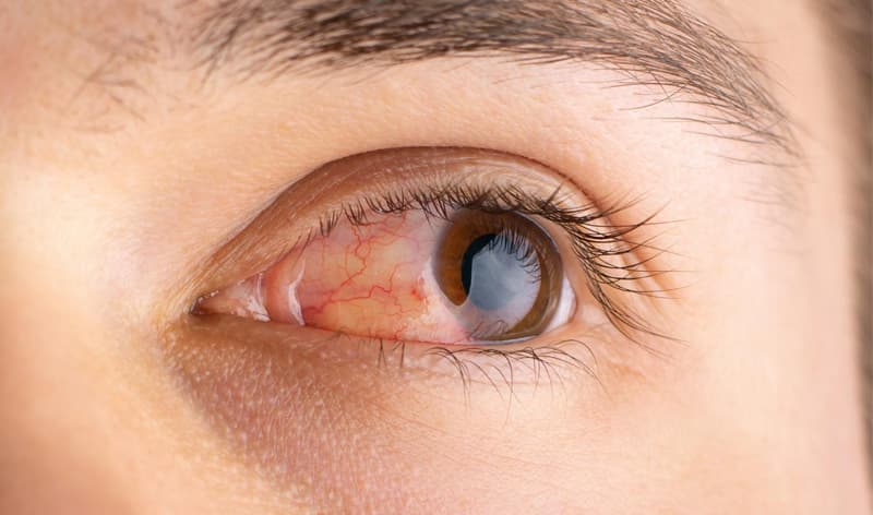
Glaucoma diagnosed through a comprehensive eye examination that measures intraocular pressure, evaluates the optic nerve for damage, and assesses peripheral vision loss. Ophthalmologists utilize a combination of tonometry, optical coherence tomography (OCT), and visual field testing to confirm the presence of the disease. This multi-test approach ensures accurate detection, even in cases of normal-tension glaucoma where eye pressure remains within standard limits, allowing for timely intervention and ongoing monitoring to prevent irreversible vision loss.
What Does a Comprehensive Glaucoma Eye Exam Feel Like for the Patient?
A comprehensive glaucoma evaluation involves a series of non-invasive, comfortable procedures designed to measure eye pressure and map the physical structures of the eye. Patients typically experience minimal discomfort, as numbing drops are applied before any instruments touch the eye surface during an eye pressure test for glaucoma. The process takes roughly 45 to 60 minutes, providing immediate insights into ocular health without requiring significant recovery time or causing prolonged blurry vision.
What Are the Key Tests Used to Diagnose Glaucoma?
Ophthalmologists rely on five primary diagnostic tools to evaluate fluid dynamics and nerve integrity within the eye. Tonometry measures intraocular pressure by gently flattening the cornea. Pachymetry measures corneal thickness , which directly influences pressure readings. An OCT scan for glaucoma captures high-resolution cross-sectional images of the optic nerve head and retinal nerve fiber layer, identifying microscopic structural damage. A visual field test for glaucoma maps the patient’s peripheral vision to detect functional blind spots. Finally, a gonioscopy test examines the drainage angle of the eye to determine the physical pathways of fluid outflow.
Why Are Multiple Tests Like OCT and Visual Field Tests Needed to Confirm a Glaucoma Diagnosis?
Relying on a single metric, such as eye pressure, is insufficient for identifying optic neuropathy because structural damage often precedes functional vision loss. An OCT scan detects physical thinning of the nerve fibers, while a visual field test measures actual vision impairment. Combining these evaluations allows specialists to understand what are the differences in diagnosing open-angle versus closed-angle glaucoma and establishes a reliable baseline for future comparison. What are the earliest signs an eye doctor looks for during a glaucoma evaluation? Clinicians search for optic disc cupping and localized nerve fiber layer defects before the patient notices any changes in their sight.
How Is Normal-Tension Glaucoma Diagnosed If Eye Pressure Readings Are Normal?
Normal-tension glaucoma occurs when optic nerve damage and vision loss progress despite intraocular pressure remaining within the statistically average range of 12 to 22 mm Hg. Diagnosis in these cases relies entirely on structural and functional assessments rather than tonometry. Specialists analyze OCT imaging for nerve thinning and conduct visual field mapping to detect characteristic peripheral defects. Blood flow evaluations and detailed medical history reviews are also necessary to rule out other vascular or neurological conditions causing the nerve damage.
How Do Glaucoma Diagnostic Tests Compare?
| Test Name | Key Feature | Best For Detecting | Patient Comfort Rating (1-5) |
|---|---|---|---|
| Tonometry | Fluid pressure measurement | Elevated intraocular pressure | 4/5 |
| OCT Scan | High-resolution laser imaging | Microscopic optic nerve damage | 5/5 |
| Visual Field Test | Light response mapping | Peripheral vision blind spots | 3/5 |
| Gonioscopy | Mirrored contact lens | Closed drainage angles | 3/5 |
| Pachymetry | Ultrasound wave measurement | Central corneal thickness | 4/5 |
What Are the Clinical Thresholds for Glaucoma Screening and Diagnosis?
Clinical evaluation for a glaucoma diagnosis follows a strict diagnostic algorithm based on age, intraocular pressure (IOP), and structural findings.
Need a comprehensive evaluation? Schedule a consultation with a glaucoma specialist in Vadodara to assess your ocular health and establish a personalized monitoring plan.
Who Should Undergo Glaucoma Screening and How Often?
Routine ocular evaluations establish early detection baselines, particularly for individuals with specific genetic or demographic risk factors. How often should I be tested for glaucoma if I have a family history of the disease? Individuals over age 40 with a first-degree relative diagnosed with the condition must undergo comprehensive screening every 1 to 2 years. Patients of African, Hispanic, or Asian descent, as well as those with diabetes or high myopia, should initiate baseline testing by age 35 to map their optic nerve structure before any potential disease onset.
What Happens After a Glaucoma Diagnosis to Monitor Its Progression Over Time?
Post-diagnosis management focuses on lowering intraocular pressure to a target level that prevents further optic nerve degradation. Specialists establish a target pressure typically 20% to 30% below the baseline measurement. Patients receive prescription eye drops, laser therapy (like SLT), or surgical interventions depending on the severity. To accurately track the disease, patients undergo repeat OCT scans and visual field tests every 6 to 12 months, allowing the clinician to adjust treatment protocols if the condition shows signs of advancement.
If you are experiencing vision changes or are due for an annual exam, contact a local eye care center to schedule a full diagnostic workup and protect your long-term vision.
Frequently Asked Questions
Modern ophthalmic clinics utilize DICOM (Digital Imaging and Communications in Medicine) standards to automatically transfer high-resolution OCT scans directly from the imaging device into the patient s electronic health record. This seamless integration allows the specialist to overlay historical scans with new images, calculating precise micrometer changes in the retinal nerve fiber layer over time.
A comprehensive glaucoma evaluation typically ranges from $150 to $400 without insurance, depending on the clinic location and the specific diagnostic instruments utilized. Health insurance plans generally cover these diagnostic tests when ordered by a physician to evaluate suspected disease, reducing out-of-pocket patient costs to standard specialist copays.
A visual field analyzer requires the patient to look into a bowl-shaped perimeter and press a button whenever they perceive a flash of light. The machine systematically presents lights of varying intensities across different quadrants of the visual field, generating a topographical map that highlights exact areas where the optic nerve is failing to transmit visual signals to the brain.
An isolated tonometry reading cannot definitively rule out the disease because up to one-third of patients with optic nerve damage have normal-tension glaucoma. Intraocular pressure fluctuates throughout the day, meaning a single normal reading might miss dangerous pressure spikes occurring at night or early in the morning.
Diagnosing closed-angle variants requires a gonioscopy test, where a specialized mirrored contact lens is placed on the eye to directly visualize the iridocorneal angle. If the ophthalmologist observes that the iris is physically blocking the trabecular meshwork drainage system, it indicates closed-angle disease, which often requires immediate laser or surgical intervention to prevent rapid vision loss.