
Medically reviewed by Dr. Meeta Mungale, MS (Ophthalmology), DNB · Cornea Fellowship, LVPEI · Gujarat Medical Council Reg. No. G-39262 · Reviewed on 3 October 2026
Published 29 September 2026 · Updated 29 September 2026
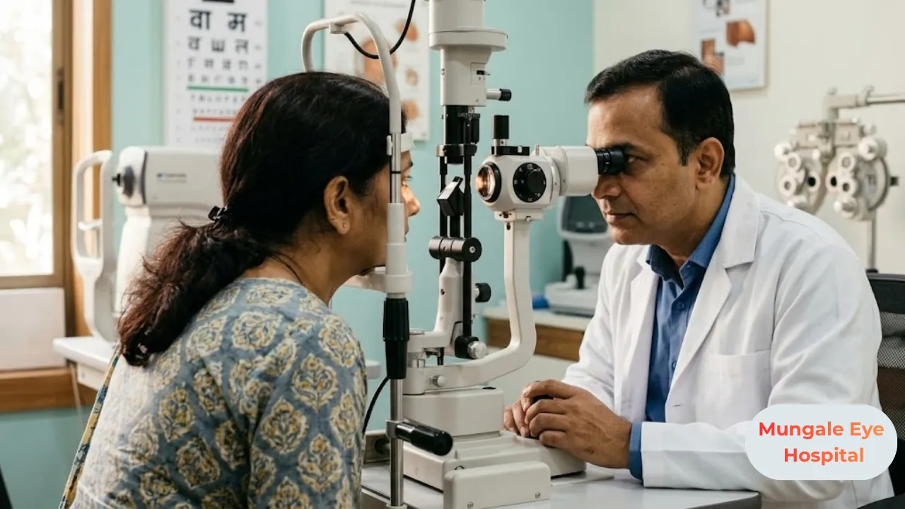
A pterygium (said tuh-RIJ-ee-um) is a pink, fleshy growth that starts on the white of the eye and slowly spreads onto the cornea, the clear dome at the front of the eye. It is not cancer and it is not an infection. Most cases are linked to years of sun, dust and wind. A small pterygium usually needs only eye drops, sunglasses and regular checks, while surgery is kept for growths that affect vision or keep flaring up.
People usually spot it in the mirror. Sometimes a family member points it out. Either way, the first reaction is often worry, and that is understandable when something new appears on your eye.
It is still worth having it looked at. A pterygium can gradually change the shape of the cornea, and left alone for years, it may start to blur your sight. Below, we explain why it forms, how doctors grade it, how the choice between drops and surgery is made, and what you can do to stop it returning after removal.
Quick Answer
A pterygium is a benign growth of the conjunctiva, the thin clear skin that covers the white of the eye. It usually begins on the side closest to the nose and creeps toward the centre of the cornea.
Long-term exposure to ultraviolet (UV) light is the biggest risk factor. Dust, heat, wind and dry air add to it.
Small pterygia are managed with lubricating drops, sun protection and monitoring. When the growth threatens vision, causes astigmatism or stays inflamed, it is removed surgically, and the area is usually covered with a thin graft of the patient's own healthy tissue to lower the chance of it growing back.
Why a Pterygium Is Worth Taking Seriously
For some people it is only a cosmetic nuisance. For others, it slowly affects how well they see.
As the growth moves onto the cornea, it tugs on the surface and flattens it in one direction. That uneven curve is called astigmatism, and it makes things look blurred or slightly stretched at every distance. Patients often say their glasses "stopped feeling right," or that the prescription keeps changing between visits.
If the pterygium keeps going and reaches the pupil, it blocks light directly. Removing it at that point is a bigger job, and a faint haze can remain on the cornea afterwards.
There is one more reason it matters. If you are planning cataract surgery or LASIK, a pterygium can throw off the measurements surgeons rely on, so it often needs attention first.
What Exactly Is a Pterygium?
The name comes from the Greek word for "wing," and the shape fits. It is broad where it sits on the white of the eye and narrows to a point, called the head, that faces the centre of the cornea.
The tissue contains fibres and small blood vessels. That is why it looks pink and turns redder when irritated.
A few things tend to reassure patients straight away. It is not cancer and does not become cancer the way many people fear. You cannot catch it or give it to someone else. It can show up in one eye or both, and it usually grows slowly, over months or years. Some stop growing on their own and stay the same for a long time.
You may hear it called "surfer's eye." The nickname comes from people who spend hours in sun and sea wind, but you do not need to be anywhere near a beach. Everyday outdoor exposure in a hot, bright, dusty place is plenty.
What Causes Pterygium?
There is no single cause, though the main triggers are well known.
Is the sun really the main cause?
Yes, as far as current understanding goes. Years of UV light reaching the eye seem to damage cells at the limbus, the narrow border where the white of the eye meets the cornea. Once that border is weakened, conjunctival tissue can grow across it.
UV light also bounces off sand, water, concrete and pale roads. So your eyes can take in a fair amount even when you are not looking anywhere near the sun.
Do dust and wind make it worse?
They do. Dust, smoke, wind and dry air keep irritating the surface of the eye, day after day, and that constant irritation seems to encourage inflammation and growth. In Vadodara, bright sun, dusty roads and daily two-wheeler travel often come together, which puts the eyes under all three kinds of stress at once.
Who tends to get it?
Pterygium is more common in people who work outdoors, such as farmers, construction workers, traffic police, delivery riders and field sales staff. Regular two-wheeler riders without protective glasses are also at higher risk, as are people who already struggle with dry, irritated eyes. It sometimes runs in families, which suggests genes may play a part.
It is uncommon in children. The risk builds with the number of years spent outdoors without eye protection.
Symptoms: How Do You Know It Is a Pterygium?
Plenty of small pterygia cause no trouble at all. People only notice the pink patch.
When symptoms do appear, the most common ones are redness that comes and goes, a gritty or sandy feeling, burning, itching and watering. The redness often gets worse after a day in the sun, a dusty ride or long hours at a screen. Contact lens wearers may find their lenses suddenly uncomfortable.
Blurred or distorted vision usually comes later, once astigmatism has set in or the growth is nearing the pupil.
A flare-up can make the eye look quite red and angry. That alone is rarely a sign of anything serious, but it should be checked so the diagnosis is confirmed and the inflammation is settled with the right treatment.
When should you book an appointment?
Any new growth on the white of your eye deserves one proper examination, even if it causes no discomfort. It is the only way to be sure it is a pterygium and not another kind of surface growth.
Try to be seen sooner if the patch seems to be getting bigger, your vision has become blurry, the redness keeps coming back despite lubricating drops, or you are planning cataract surgery, LASIK or a contact lens fitting.
When is it urgent?
Please seek same-day eye care if you have:
A few uncommon surface conditions can look a lot like pterygium and need different care. An unusual growth should be seen by an ophthalmologist, not watched at home.
Stages of Pterygium
Doctors describe a pterygium by how far it has travelled across the cornea and by how active it looks. Several grading systems are in use. One common way of grading by extent is shown here.
| Grade | How far it has grown | What it usually means |
|---|---|---|
| Grade 1 | Up to the limbus (edge of the cornea) | Often no symptoms; monitoring and sun protection |
| Grade 2 | Onto the cornea, well short of the pupil | May irritate or cause mild astigmatism; checked regularly |
| Grade 3 | To the edge of the pupil | Blur and astigmatism more likely; surgery often discussed |
| Grade 4 | Across the pupil | Can block vision; surgery usually advised |
Appearance matters too. A thin, pale pterygium with fine vessels is usually quiet and slow. A thick, raised, red one with obvious vessels is more active, more likely to grow, and more likely to come back after surgery.
Your doctor may photograph the eye at each visit. Placing those photos side by side over a year or two is one of the simplest and most reliable ways to know whether it is really growing.
How a Pterygium Affects Your Vision
It works in two ways.
First, it reshapes the cornea. The pull of the growth flattens one part of the corneal surface, producing astigmatism. You might not notice much at first, but a scan can pick it up early.
Second, once the head of the growth reaches the pupil, it simply gets in the way of light.
The scan used to measure astigmatism is called corneal topography. It is painless and takes a few minutes, producing a colour map of the corneal curve. This is partly why surgeons sometimes suggest removal before symptoms feel severe. An earlier removal often lets the cornea regain much of its normal shape. A very advanced pterygium may leave some irregularity or haze behind.
How Is Pterygium Diagnosed?
Usually in a single visit. At Mungale Eye Hospital, the assessment normally starts with a vision chart and a glasses check, which shows whether your sight has been affected.
The doctor then looks at the eye under a slit lamp, a microscope with a narrow, bright beam that shows the growth, its blood vessels and how far it has spread. Corneal topography maps any astigmatism. In some cases an anterior segment OCT is added. This is a quick, no-touch scan that shows the front layers of the eye in fine detail and helps judge the depth of the growth.
Photographs are taken as a baseline for future comparison.
If anything about the growth looks unusual, the doctor may advise removing it and sending the tissue to a laboratory to confirm exactly what it is.
Treatment: Eye Drops or Surgery?
The choice depends on the size of the pterygium, how much it bothers you, whether it is affecting vision, and whether other eye surgery is planned. You and your ophthalmologist make that decision together.
What eye drops can and cannot do
Drops do not shrink or dissolve a pterygium. They make the eye more comfortable and calm inflammation, and for many people that is enough.
Lubricating drops or gels (artificial tears) ease dryness and grittiness. During a red, irritated flare-up, your doctor may prescribe a short course of anti-inflammatory drops. Steroid drops should only ever be used under an eye doctor's supervision. Used on their own for long periods, they can raise eye pressure and cause other problems.
Alongside drops, sun protection does real work. UV-blocking sunglasses, a cap or wide-brimmed hat, and protective glasses while riding or working in dust all reduce the daily strain on the eye. Regular reviews then tell you whether the growth is stable.
When does surgery make sense?
Surgery is usually suggested when the pterygium is heading toward the pupil or already causing astigmatism that blurs vision. Persistent redness and discomfort despite drops is another common reason. So is trouble wearing contact lenses, or an upcoming cataract or refractive procedure that needs accurate corneal measurements.
A growth that looks atypical may be removed so the tissue can be examined. Rarely, an advanced or recurrent pterygium restricts eye movement and causes double vision, which also calls for surgery.
Some people want it removed mainly because of how it looks. That is a fair concern and worth discussing openly, keeping in mind that recurrence risk applies here too.
| Approach | What it does | Usually considered for |
|---|---|---|
| Monitoring and sun protection | Tracks growth, reduces UV and dust | Small, quiet pterygium not affecting vision |
| Lubricating drops | Eases dryness and grittiness | Mild irritation at any stage |
| Supervised anti-inflammatory drops | Settles redness during flare-ups | Short-term use for inflamed pterygium |
| Excision with conjunctival autograft | Removes the growth, covers the area with your own healthy tissue | Growing, symptomatic or vision-affecting pterygium |
| Excision with amniotic membrane graft | Removes the growth, covers the area with a processed biological membrane | Large or recurrent pterygium, or when your own tissue needs to be saved |
What Happens During Pterygium Surgery?
It is a day-care procedure done under local anaesthesia. You are awake throughout, but the eye is numb. Most people go home a short while after.
Getting the eye ready
The area around the eye is cleaned. Numbing drops, sometimes with a small local injection, take away sensation. A light clip keeps the lids open, so you do not have to think about blinking.
Removing the growth
The surgeon lifts the head of the pterygium gently off the cornea and takes away the abnormal tissue from the white of the eye. The corneal surface is then smoothed.
Covering the area with a graft
In older techniques, the white of the eye was simply left bare after removal. Regrowth was more common with that approach, so today the area is usually covered.
The most common option is a conjunctival autograft. A thin piece of healthy conjunctiva is taken from your own eye, generally from under the upper lid, where it heals well and stays out of sight. It is placed over the treated area. Since it is your own tissue, the body does not reject it.
An amniotic membrane graft is a specially prepared biological membrane. Surgeons may choose it for a large area, for a pterygium that has returned after earlier surgery, or when your own conjunctiva is better preserved.
The graft is held in place with fibrin glue, a tissue adhesive, or with very fine stitches. Glue tends to be more comfortable afterwards. Stitches suit certain situations better. Your surgeon will tell you which is planned and why.
For some recurrent cases, an extra medication may be applied during surgery to reduce regrowth. It has its own considerations and is decided case by case.
At Mungale Eye Hospital, pterygium removal with conjunctival autograft and amniotic membrane grafting is part of the cornea service, led by Dr. Sachin Mungale (MS, Ophthalmology) and Dr. Meeta Mungale (MS, Ophthalmology, DNB).
Recovery After Pterygium Surgery
Healing is usually uneventful, though the eye needs time. Everyone recovers a little differently, so your own surgeon's instructions come first.
In the first few days, expect grittiness, watering and mild soreness. The eye will be red, and there may be a patch of blood on the white. It looks alarming to some patients but normally clears without treatment. You may wear a pad on day one and a shield at night for a short while. Drops start straight away, typically an antibiotic and an anti-inflammatory.
Over the following weeks, discomfort fades, usually within a week or so, though some grittiness can hang around. The graft may stay a little pink for several weeks before it blends in. Anti-inflammatory drops are reduced slowly over this period. Please don't stop them early because the eye feels fine. That slow taper is one of the main things keeping regrowth in check.
If stitches were used, they either dissolve or are removed at a follow-up.
A few simple habits make a big difference. Don't rub or press the eye. Keep water, soap and shampoo out of it while bathing for as long as your doctor advises. Stay away from swimming pools, dust and smoke until you are cleared. Wear sunglasses outdoors from the very first day, and wash your hands before putting in drops.
Most people are back at a desk job within a few days, depending on comfort. Outdoor or dusty work may need a longer break, and your surgeon can advise on timing.
Keep every follow-up appointment. The early visits check that the graft is healing in place. The later ones look for any sign of the pterygium returning.
Can Pterygium Come Back After Surgery?
It can. When it does, it most often happens within the first several months, which is exactly why those follow-up visits matter.
Several things influence the risk. Grafting techniques carry a lower recurrence risk than leaving the area bare. A thick, fleshy, active pterygium is more likely to return than a thin, quiet one. Younger patients seem to have a stronger tendency toward regrowth, and a pterygium that has already come back once is more likely to come back again. Continued unprotected sun and dust exposure after surgery may also play a part.
What helps most is fairly simple. Finish every drop in the prescribed taper. Wear UV-blocking sunglasses whenever you are outside, add a cap in strong sun, and use protective glasses on a two-wheeler. Keep the eyes lubricated if they tend to feel dry. If you notice new redness or a fleshy patch at the surgery site, get it checked early.
If it does come back, you have not done anything wrong. Your ophthalmologist can talk you through further options, which may include repeat surgery with additional measures.
Risks of Pterygium Surgery
It is a commonly performed operation and serious problems are uncommon. Still, you should know what can happen.
Recurrence is the main one. Temporary redness, watering, grittiness and light sensitivity are expected. Bleeding under the conjunctiva usually clears by itself. Occasionally the graft swells or shifts and needs attention. Infection is rare but needs prompt treatment.
After removal of a large pterygium, a small amount of haze or scarring may remain on the cornea, along with some residual astigmatism that updated glasses can often correct. In some people, steroid drops raise eye pressure, which is one of the things checked at follow-up.
Your surgeon will explain which of these are more or less likely for your particular eye.
Pterygium or Pinguecula?
These two get mixed up all the time. Both are linked to sun, dust and wind, and both sit on the white of the eye. The difference comes down to one thing. A pinguecula (said pin-GWEK-yoo-luh) stays on the white of the eye. A pterygium grows onto the cornea.
| Feature | Pinguecula | Pterygium |
|---|---|---|
| Looks like | Small, raised, yellowish-white spot | Pink or red, fleshy, wing-shaped growth |
| Where it sits | On the conjunctiva, beside the cornea | Crosses onto the cornea |
| Effect on vision | Usually none | Can cause astigmatism or block vision as it grows |
| Typical symptoms | Occasional irritation or redness when inflamed | Irritation, redness, grittiness, later blur |
| Usual treatment | Lubricants, sun protection, short anti-inflammatory course if inflamed | Same early on; surgery if growing or affecting vision |
The two can sit side by side in the same eye. Only an examination tells them apart with confidence.
Cost and Insurance for Pterygium Surgery
There is no honest single price for pterygium surgery without an examination, because a few things change the final figure.
The biggest factor is the type of surgery, whether a conjunctival autograft, an amniotic membrane graft or extra steps for a recurrent pterygium. Glue or stitches, the pre-operative scans needed, post-operative medicines and follow-ups, and any laboratory examination of the removed tissue all add to the total.
Many health insurance policies in India cover pterygium surgery when it is medically needed, for instance when vision is affected or symptoms persist. Cover for purely cosmetic removal may be limited, and waiting periods can apply. Read the eye-surgery clauses in your policy, or let the hospital team check it with you.
Mungale Eye Hospital offers insurance and cashless treatment support, and the team can help you understand likely cover before you decide.
Myths We Hear Often
"It's a kind of cancer." It isn't. A pterygium is benign. An odd-looking growth still needs checking, because a few rare conditions can look similar.
"The right drops will dissolve it." Drops ease dryness and inflammation. They do not remove the tissue. Surgery is the only way to do that.
"It always comes back after surgery." Recurrence happens, but many people never see it return, especially with a graft, careful aftercare and steady sun protection.
"You can catch it from someone." You can't. It is not an infection.
"Wait until it covers your vision, then operate." Very advanced growths can leave more corneal distortion after removal. The timing should be decided with your ophthalmologist based on growth, astigmatism and symptoms.
Mistakes That Make Things Harder
The most common one is reaching for old or over-the-counter steroid drops whenever the eye turns red. They may calm it briefly, but without supervision they can raise eye pressure and hide other problems.
Skipping sunglasses is another. UV protection is one of the few measures that clearly helps with pterygium, both before and after surgery.
After surgery, some people stop their drops as soon as the eye feels better. The slow taper is there for a reason.
And if you are planning cataract surgery or LASIK, make sure your surgeon knows about the pterygium. It can affect the planning.
Looking After Your Eyes Day to Day
If you have a pterygium, or you are at risk of one, a few habits go a long way.
Get any new growth checked once, then keep to the review schedule your doctor sets. Wear UV400 or equivalent UV-blocking sunglasses outside, cloudy days included. Wraparound frames are worth considering if you ride or work outdoors, since they block light and dust from the sides as well. If you use drops often, ask whether a preservative-free option suits you. At a screen, take short breaks and remember to blink fully, which helps keep the eye surface from drying out.
A Few Practical Tips From the Clinic
If your glasses prescription keeps changing and you have a pterygium, ask for a topography scan. It will show whether the growth is causing the astigmatism.
Ask your surgeon which graft is planned and why. Knowing whether it will be an autograft or amniotic membrane helps you understand the recovery.
If you work outdoors, try to time surgery for a stretch when you can rest and avoid dust for a while.
Old photographs help too. Pictures that clearly show your eyes from a few years ago can tell your doctor how quickly the growth has changed.
Choosing Where to Get Pterygium Treatment
Pterygium surgery is part of cornea care. It helps to look for ophthalmologists with real experience in corneal and ocular surface surgery, grafting techniques such as conjunctival autograft and amniotic membrane grafting used as a matter of routine, and diagnostic tools like corneal topography and anterior segment OCT. Clear explanations of recurrence risk and aftercare, along with upfront guidance on cost and insurance, matter just as much.
Mungale Eye Hospital in Kothi (Anandpura), Vadodara, has provided eye care since 2007 and serves as a referral centre for complex cornea and glaucoma cases.
What Has Changed in Pterygium Treatment
Surgery has become more refined over the years. Covering the treated area with the patient's own conjunctiva has largely replaced the bare sclera method. Fibrin glue has made grafts more comfortable to recover from. Amniotic membrane is now a standard option for larger or recurrent growths.
Research continues into ways to lower recurrence further, including medicines aimed at the blood vessel growth seen in active pterygia. Some of these are still being studied, and your surgeon will only recommend what is appropriate and established for your eye.
Key Takeaways
Final Thoughts
Pterygium is common, usually slow and very treatable. Plenty of people live with a small one for years with nothing more than sunglasses and the occasional check-up. For others, well-timed surgery with a graft clears the growth, settles the irritation and lets the cornea recover a healthier shape.
The most useful step is simply getting it examined. One visit confirms what the growth is, shows whether it is affecting your sight, and gives you a clear plan from there. If you have noticed something on the white of your eye, or you have been told you have a pterygium and want to understand your choices, the cornea team at Mungale Eye Hospital, Vadodara, can examine your eyes and talk you through them.
This article provides general health information and is not a substitute for professional medical advice, diagnosis or treatment. Please consult a qualified ophthalmologist for an in-person examination.
FAQ SECTION
A pterygium is benign and not dangerous in itself. Over time, though, it can change the shape of the cornea and cause astigmatism, and a large one may block vision if it reaches the pupil. Regular check-ups let your ophthalmologist see whether it is growing and recommend treatment before your eyesight is noticeably affected.
It usually doesn't disappear by itself. Lubricating drops and sun protection can calm redness and irritation, which may make it look less obvious, and some pterygia stop growing and stay stable for years. If the growth needs to be removed, surgery is the only treatment that physically takes the tissue away.
The surgery is done under local anaesthesia, so the eye is numb and you should not feel pain during it. Afterwards, grittiness, watering and mild soreness are common for several days. These ease gradually, and the drops your doctor prescribes help keep the eye comfortable while it heals.
Most people feel fairly comfortable within about a week and return to desk work in a few days. Redness at the surgical site fades more slowly and can take several weeks to settle. Anti-inflammatory drops are reduced gradually over weeks, and follow-up visits continue for some months to check healing and watch for regrowth.
It can, most often within the first several months. The risk depends on the surgical technique, how active the pterygium was, your age and how much sun you get afterwards. Graft techniques lower the risk compared with older methods. Finishing your drops, wearing UV-blocking sunglasses and keeping follow-up appointments all help.
Both appear on the white of the eye and are linked to sun, dust and wind. A pinguecula is a small yellowish bump that stays beside the cornea and rarely affects vision. A pterygium is a pink, fleshy, wing-shaped growth that moves onto the cornea and can cause astigmatism or blocked vision as it grows.
Often it should, if it is large enough to distort the cornea. That distortion can affect the measurements used to choose the lens implant for cataract surgery. Removing the pterygium first and letting the cornea settle can give more accurate lens calculations. Your ophthalmologist will decide the order after examining both problems.
Many policies cover it when it is medically necessary, for example when vision is affected or symptoms persist. Cover for purely cosmetic removal may be limited, and waiting periods can apply. Check your policy's eye-surgery terms, and ask the hospital's insurance desk to confirm eligibility and cashless options before your surgery date.
Cross-cluster links for the body: Dry Eye Disease pillar (lubricants and surface irritation), Cataract Surgery pillar (pterygium removal before lens measurements), Corneal Transplant pillar (advanced corneal scarring).